
Lymphedema and Lymphatics
Home
The goal of this work is to apply novel, noninvasive magnetic resonance imaging (MRI) methods for visualizing lymphatic circulation dysfunction to test fundamental hypotheses about lymphedema risk factors and therapies. Breast cancer treatment-related lymphedema (BCRL) arises secondary to surgical axillary lymph node (LN) dissection and irritation, and is a chronic and lifelong condition affecting a high 21.4% of patients receiving common breast cancer therapies. Reducing condition onset and improving management represent major unmet clinical needs for these 50,000 - 80,000 new patients per year, and emerging efforts focus on improving quality of life through more informed LN dissection and biopsy decisions, optimizing post-surgical complex decongestive therapy (CDT), and exploring novel pharmacological and surgical procedures. However, fundamental gaps in our knowledge persist regarding optimized implementation of these therapies and details of the physiological changes they elicit. The major underlying limitation is that there is a shortage of imaging methods available that can be used to evaluate lymphatic function directly, and there is currently no consensus regarding effective outcome measures for therapeutic efficacy evaluation.
Our manuscripts relevant to this work in lymphatics and cancer:
Since approximately 2013, we have worked on developing methods to assess lymphatic collector structure and function before and after cancer therapies. Specifically, BCRL is a chronic, debilitating disease caused by lymphatic flow obstruction and lymphedema secondary to mastectomy with radiation therapy has been reported to occur in 20-30% of breast cancer survivors following these therapies. However, owing to a lack of methodology for sensitively identifying lymphatic system compromise, there are important gaps in our knowledge regarding which patients are at highest risk and how and when therapies should be applied to minimize impairment. In this work, novel and noninvasive magnetic resonance imaging approaches for measuring lymphatic dysfunction are applied to improve procedures for preventing, predicting, and treating BCRL.
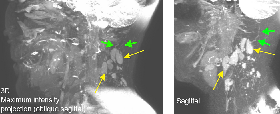
Our initial trials in BCRL have ended and are open for data analysis only. Ongoing extensions of this work focus on evaluating lymphatic tissue structure and function in the neck, and specifically, how such cervical lymphatic changes relate to retention of cerebral peptides in patients with neurodegenerative proteinopathies.